Upper GI
Bilary Cancer
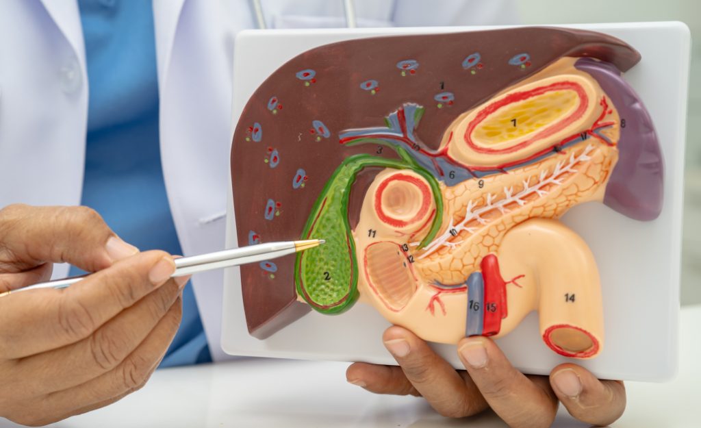
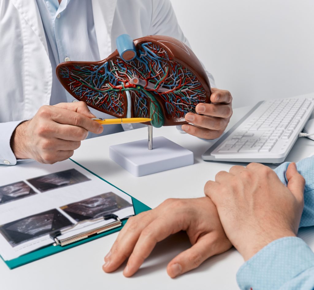
Liver Lesions: Patient Information
Liver lesions are abnormal growths or areas of damage in the liver. They can be benign (non-cancerous) or malignant (cancerous).
The liver is a vital organ that plays a crucial role in detoxifying the body, producing bile for digestion, and storing nutrients.
Types of Liver Lesions
- Benign Liver Lesions:
- Haemangioma: A common, benign blood vessel tumour.
- Hepatic Adenoma: A rare, benign tumour often associated with the use of oral contraceptives or anabolic steroids.
- Focal Nodular Hyperplasia (FNH): A benign growth often found in young women.
- Cysts: Fluid-filled sacs that are usually benign.
- Malignant Liver Lesions:
- Hepatocellular Carcinoma (HCC): The most common type of primary liver cancer, often associated with chronic liver diseases like hepatitis and cirrhosis.
- Cholangiocarcinoma: Cancer that forms in the bile ducts.
- Metastatic Liver Cancer: Cancer that has spread to the liver from other parts of the body, such as the colon, breast, or lung.
Causes and Risk Factors
- Chronic Liver Diseases: Hepatitis B and C, cirrhosis, non-alcoholic fatty liver disease (NAFLD).
- Genetic Conditions: Hemochromatosis, Wilson’s disease.
- Lifestyle Factors: Alcohol abuse, obesity.
- Medications: Long-term use of oral contraceptives or anabolic steroids.
- Family History: Genetic predisposition to liver conditions.
Symptoms
- Asymptomatic: Many liver lesions are found incidentally during imaging tests for other conditions.
- Abdominal Pain: Especially in the upper right quadrant.
- Swelling: Abdominal swelling or a palpable mass.
- Jaundice: Yellowing of the skin and eyes.
- Unexplained Weight Loss: Significant weight loss without trying.
- Fatigue: Persistent tiredness and weakness.
- Nausea and Vomiting: Especially if the lesion affects liver function.

Diagnosis
- Imaging Tests:
- Ultrasound: Often the first imaging test to detect liver lesions.
- CT Scan: Provides detailed cross-sectional images of the liver.
- MRI: Offers detailed images and helps distinguish between different types of lesions.
- PET Scan: Used to detect cancer spread.
- Blood Tests:
- Liver Function Tests (LFTs): Assess the liver’s overall function.
- Tumour Markers: Alpha-fetoprotein (AFP) for HCC, CA 19-9 for cholangiocarcinoma, CEA for metastatic colon cancer.
- Biopsy: A sample of liver tissue is taken for microscopic examination to determine if a lesion is benign or malignant.
Treatment Options
Benign Lesions
- Observation: Many benign lesions do not require treatment and are monitored for changes.
- Surgery: If the lesion causes symptoms or complications, surgical removal may be necessary.
Malignant Lesions
- Surgery: Surgical resection of the tumour, often the best option for early-stage cancer.
- Liver Transplant: For certain cases of HCC or other extensive liver diseases.
- Ablation Therapy: Techniques like radiofrequency ablation (RFA) or microwave ablation to destroy cancer cells.
- Embolization: Transarterial chemoembolization (TACE) or radioembolization to block blood supply to the tumour.
- Radiation Therapy: External beam radiation or selective internal radiation therapy (SIRT).
- Chemotherapy: Systemic chemotherapy or targeted drugs like sorafenib for advanced HCC.
- Immunotherapy: Drugs that help the immune system attack cancer cells, such as nivolumab.
Trusted for care
Dr Wang and his team are respected and trusted for the care provided to their patients

Follow-Up Care
- Regular Monitoring: Follow-up imaging and blood tests to monitor for recurrence or new lesions.
- Liver Health: Managing underlying liver conditions and maintaining liver health.
- Lifestyle Changes: Avoiding alcohol, maintaining a healthy weight, and following a balanced diet.
When to Seek Medical Attention
- New or worsening symptoms such as severe abdominal pain, jaundice, or unexplained weight loss.
- Symptoms of liver dysfunction, such as confusion or easy bruising.
- Signs of infection at a biopsy site, including fever, redness, or swelling.

Other Upper GI Conditions
Pancreatic cancer begins in the tissues of the pancreas, an organ located behind the stomach…
Also known as stomach cancer, is a disease in which malignant (cancer) cells form in the lining…
Medical procedure used to examine the upper part of the GI Tract, colon and rectum for abnormalities….
Also known as stomach cancer, is a disease in which malignant (cancer) cells form in the lining…